CT Scans Cause Brain Cancer, Leukemia
According to a new study funded by the US National Cancer Institute and the UK’s Department of Health, having CT scans during ones youth can triple the risk of brain cancer and leukemia later on.
This is forcing doctors, hospitals and patients to question the need for CT scans in some cases.
In this article
Large study testing CT scans
The research was conducted among 178,604 people, including those who received a CT scan before the age of 22 years old between 1985 and 2002. The researchers then analyzed the data for cancer occurrences and deaths from cancer between 1985 and 2008. The follow-up period testing began two years from the patient’s first CT scan.
The analysis found that those receiving a CT scan or scans with more than 30 mGy of radiation dosage had 318% (over three times) greater incidence of leukemia, and those receiving more than 50 mGy of CT-scan radiation had a 282% (2.8 times) greater incidence in brain cancer compared to those who did not receive a CT-scan, or received a very low dosage scan.
Previous research has determined that children are more sensitive to radiation than adults.
What is a CT scan?
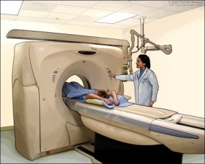
A CT scan uses an imaging technique called computed tomography. It is one of the highest sources of ionizing radiation exposure within the health care industry. The use of CT scans has increased dramatically over the past decade, and many health experts have stated that it is often an unnecessary procedure. It is sometimes ordered even if there is confidence in the diagnosis, just to cover the potential for a malpractice suit. In other words, hospitals and physicians sometimes order CT scans for patients as a type of insurance policy.
The researchers concluded that, “use of CT scans in children to deliver cumulative doses of about 50 mGy might almost triple the risk of leukaemia and doses of about 60 mGy might triple the risk of brain cancer.”
CT-scans are often regional scans. Radiation dosage will depend upon the surface area scanned, as well as upon the image resolution and image quality of the scan.
Other research has established that ones combined radiation exposure load or dosage – a combination of ionizing radiation exposure from various sources – is a more appropriate predictor for cancer incidence from radiation.
This is a far larger link than found from cell phone use. Although long-term cell phone use is linked to brain cancer too.
Needless CT scans done by U.S. hospitals
Researchers have found that hospitals are ordering more CT Scans than necessary. Medicare records and interviews with medical experts has determined that hospital physicians are ordering double CT scans when they are, according to radiologists, only rarely required.
According to a New York Times report, hospitals ordered two CT scans for 80 percent of their Medicare patients. The average rate of double-CT Scans is typically 1%, according to the report. Teaching hospitals at medical schools, meanwhile, rarely if ever order double CT scans. Other research also finds some hospitals conducting unnecessary tests and procedures.
The growth in CT scans over the past decades has been astronomical. About sixty-two million CT scans are now given a year in the U.S., as opposed to about three million per year in 1980. A study published in the New England Journal of Medicine showed that a third of CT scans are unnecessary. The authors also estimated that between one and two percent of all cancers are caused by CT scan radiation exposure.
A CT scan will can render a dose of about 10 mSv—making a CT scan between a hundred and a thousand times the dose of an x-ray. In contrast, the maximum radiation a nuclear electricity generating plant will emit at the perimeter fence is about .05 mSv per year. A set of dental x-rays will render a dose of about .05-.1 mSv.
REFERENCES:
Pearce MS, Salotti JA, Little MP, McHugh K, Lee C, Kim KP, Howe NL, Ronckers CM, Rajaraman P, Craft AW, Parker L, de González AB. Radiation exposure from CT scans in childhood and subsequent risk of leukaemia and brain tumours: a retrospective cohort study. Lancet, 7 June 2012. doi:10.1016/S0140-6736(12)60815-0
Brenner DJ, Hall EJ. Computed tomography–an increasing source of radiation exposure. N Engl J Med. 2007 Nov 29;357(22):2277-84.
Adams C. Electromagnetic Health: Making Sense of the Research and Practical Solutions for Electromagnetic Fields (EMF) and Radio Frequencies (RF) Logical Books, 2015.

